
General education only; not a substitute for examination or diagnosis.
What this area covers
The ear, nose and sinuses lie very close to the skull base, the eye socket (orbit) and the membranes around the brain. Only a thin plate of bone separates the middle ear and mastoid from the brain, and the sinus walls border the eye socket and the front of the skull base. For this reason a prolonged or severe infection can, although rarely, spread to neighbouring structures.
Complex ENT disease covers such spread, disease that persists despite previous surgery and difficult infections in people with weakened immunity or after radiotherapy to the head and neck. These patients are often managed by a multidisciplinary team including neurosurgery, ophthalmology, infectious diseases, radiology and neurology.
- Cholesteatoma and its complications: mastoiditis, facial palsy, a bony defect opening into the inner ear (labyrinthine fistula) and intracranial complications such as meningitis, brain abscess, abscess around the brain membranes or a clot in the venous sinus behind the ear (sigmoid sinus thrombosis).
- Spread of sinusitis to the tissues around the eye and into the orbit, and intracranial complications such as meningitis or abscess.
- Revision surgery for persistent or recurrent disease in ears or sinuses that have already been operated on.
- Leakage of cerebrospinal fluid from the nose (CSF rhinorrhoea).
- Persistent or recurrent ear and sinus infections in people with diabetes, immunosuppressive treatment or previous radiotherapy; for example necrotising (malignant) otitis externa spreading from the ear canal to the skull base, and invasive fungal sinusitis in severely immunocompromised patients.
Symptoms
- Foul-smelling, usually one-sided ear discharge that does not clear with treatment, with progressive hearing loss.
- Pain, redness or swelling behind the ear, or the ear being pushed forward.
- Weakness on one side of the face, inability to close the eye or drooping at the corner of the mouth.
- Dizziness; vertigo brought on by pressing on the ear or blowing the nose may suggest a labyrinthine fistula.
- During sinusitis: swelling of the eyelids, red eye, a bulging eye, double vision or pain on moving the eye.
- Severe headache, neck stiffness, high fever, confusion, swelling over the forehead or seizures.
- Clear, watery discharge from one side of the nose that increases on leaning forward; it may follow head injury or nose or sinus surgery.
When to see a doctor
Situations that need urgent care
- Swelling around the eye, a bulging eye, double vision, restricted eye movement or reduced vision during sinusitis or a nasal infection.
- Severe headache, neck stiffness, altered consciousness, seizures or limb weakness with an ear or sinus infection: call emergency services (112).
- New facial palsy in someone with ear disease.
- Rapidly increasing painful swelling behind the ear with high fever.
- Clear fluid from the nose after head injury or nasal surgery together with fever and headache.
- Rapidly worsening facial pain, black crusting in the nose or severe ear pain in a person with weak immunity or diabetes.
Situations for a planned appointment
- Ear discharge that lasts for weeks or keeps coming back.
- Symptoms that continue or return despite previous ear or sinus surgery.
- Long-standing one-sided, watery nasal discharge.
- Ear blockage, hearing loss or chronic sinus symptoms after head and neck radiotherapy.
Some of these symptoms can also have simpler causes. A definite diagnosis and treatment decision is made after examination and the necessary tests.
How it is diagnosed
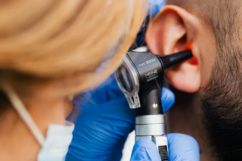
- History and examination: examination of the eardrum and mastoid cavity with an ear microscope, assessment of the nose and sinus openings with a rigid or flexible endoscope, and checks of facial nerve function and eye movements.
- Hearing tests: pure-tone audiometry and tympanometry, documenting hearing before surgery when cholesteatoma or inner-ear involvement is present.
- Imaging: CT of the temporal bone or sinuses shows bony anatomy and spread; contrast-enhanced CT and MRI are added when an intracranial or orbital complication is suspected. Diffusion-weighted MRI helps detect residual or recurrent cholesteatoma after surgery.
- When a CSF leak is suspected, beta-2 transferrin testing of the nasal fluid is the most reliable confirmatory test; high-resolution CT is the first choice for locating the leak, with MR cisternography when needed.
- Cultures and blood tests: samples from discharge or abscess identify the organism and the right antibiotic, and biopsy is taken from suspicious tissue.
- Ophthalmology, neurosurgery, neurology and infectious diseases consultations when needed.
Treatment options
Close monitoring and supportive care
Spread of sinusitis to the eye is classically described in stages (the Chandler classification): eyelid swelling (preseptal cellulitis), inflammation of the orbital tissues (orbital cellulitis), subperiosteal abscess, orbital abscess and, most seriously, cavernous sinus thrombosis.
Some early infections around the eye and limited complications can be controlled with medicines under close monitoring in hospital. Vision, eye movements and level of consciousness are checked at regular intervals, and the plan is changed quickly if things worsen.
Medicines
- Complicated infections are usually treated with intravenous antibiotics in hospital, adjusted according to culture results.
- For chronic ear discharge, cleaning the ear and ear drops are the basis of treatment.
- For invasive fungal infection, antifungal medicines, surgical debridement and correction of the underlying immune problem are planned together.
Surgery
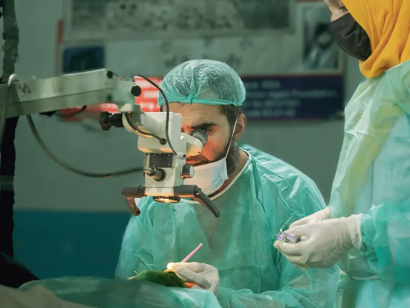
- Surgery is the main treatment for cholesteatoma; it does not disappear with medicines. Diseased tissue is removed by mastoidectomy, and in suitable patients hearing is reconstructed in the same operation or a later one.
- If an abscess forms in the orbit, vision deteriorates or there is no response to medicines, drainage by endoscopic sinus surgery is considered.
- For intracranial abscesses, surgery on the source in the ear or sinus is planned together with neurosurgery.
- For CSF leaks, endoscopic repair through the nose may be performed in selected patients; vascularised tissue flaps may be preferred for larger defects.
- For revision surgery, previous operative records and current imaging are reviewed in detail, and planning is more careful because the anatomy has changed.
Before and after surgery
Before surgery, imaging, hearing tests, blood tests and an anaesthetic assessment are completed. Good control of diabetes and other conditions is important for healing. Blood thinners should only be stopped or adjusted on the advice of the treating physician.
Length of hospital stay and recovery depend on the type of operation and the severity of the complication. After ear surgery keeping the ear dry, and after nose and sinus surgery avoiding forceful nose blowing and heavy lifting, are usually advised.
- High fever, severe headache, neck stiffness, clear fluid from the nose, changes in vision or facial weakness after surgery need immediate medical attention.
Follow-up
- Because cholesteatoma can recur, regular ear examination is needed for years after surgery; in some patients diffusion-weighted MRI or a planned second-look operation is used.
- After a sinus complication, healing is monitored with endoscopic checks and imaging when needed.
- After CSF leak repair, patients are monitored for recurrent nasal fluid and headache.
- In people who have had radiotherapy or have weak immunity, new symptoms should be assessed without delay.
Frequently asked questions
Sources
The general information on this page is based on the guideline and institutional sources below. Individual diagnosis and treatment decisions require an examination.
- NHS — Cholesteatoma
- StatPearls (NCBI Bookshelf) — Middle Ear Cholesteatoma
- NICE NG79 — Sinusitis (acute): recommendations including hospital referral criteria
- Chandler JR et al. The pathogenesis of orbital complications in acute sinusitis. Laryngoscope, 1970
- Oakley GM et al. Diagnosis of cerebrospinal fluid rhinorrhea: an evidence-based review with recommendations. Int Forum Allergy Rhinol, 2016
- Oakley GM et al. Management of cerebrospinal fluid rhinorrhea: an evidence-based review with recommendations. Int Forum Allergy Rhinol, 2016