
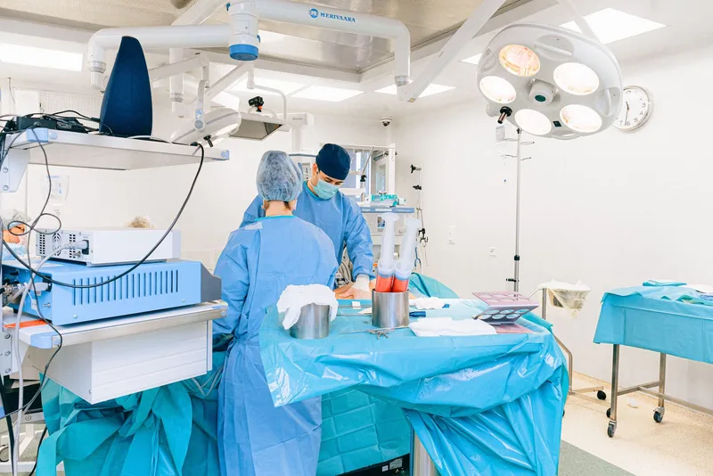
General education only; not a substitute for examination or diagnosis.
What head and neck cancers are
Head and neck cancers are tumours that arise in the oral cavity, the pharynx (throat), the larynx (voice box), the nasal cavity and sinuses, and the salivary glands. Most cancers in this region are squamous cell carcinomas, which start in the cells lining the inside of the mouth and throat. Thyroid, brain, eye and skin cancers are usually not classified in this group.
- Oral cavity: the lips, the front two-thirds of the tongue, the gums, the inner cheek, the floor of the mouth and the hard palate.
- Oropharynx: the tonsils, the base of the tongue, the soft palate and the back wall of the throat; a large share of tumours here are linked to HPV (human papillomavirus).
- Larynx: the area containing the vocal cords; in vocal cord tumours hoarseness often appears early.
- Hypopharynx: the part of the throat behind and beside the larynx that leads into the oesophagus.
- Nasopharynx: the uppermost part of the throat behind the nose; cancer in this area is associated with EBV (Epstein–Barr virus).
- The nasal cavity and paranasal sinuses (the sinonasal region), and the major and minor salivary glands.
Tobacco (including cigarettes, waterpipe and smokeless tobacco) and alcohol are the most important risk factors, and using both raises the risk further. According to the US National Cancer Institute (NCI), about three-quarters of oropharyngeal cancers are linked to chronic HPV infection. EBV is a risk factor for nasopharyngeal cancer; other risk factors include betel quid (paan) chewing, some occupational exposures such as wood dust, and previous radiotherapy to this region.
Symptoms
Most of the complaints below can also be caused by harmless conditions such as infections. What matters is that the complaint does not go away and lasts longer than about three weeks.
- A mouth ulcer or lip sore that has not healed after three weeks.
- A red (erythroplakia) or white (leukoplakia) patch inside the mouth.
- Persistent hoarseness or voice change; the American Academy of Otolaryngology (AAO-HNSF) guideline recommends examining the larynx when hoarseness has not improved within four weeks.
- A lump in the neck that does not go away or is getting bigger.
- Difficulty swallowing (dysphagia), pain on swallowing (odynophagia) or a feeling of food sticking in the throat.
- One-sided ear pain; if the ear itself looks normal, the pain may be referred from the throat or tongue base.
- One-sided nasal blockage, recurrent nosebleeds or blood-stained nasal discharge.
- Blood in the saliva, unexplained weight loss or loosening teeth.
When to see a doctor
Situations that need emergency care
- Shortness of breath or a noisy, high-pitched sound when breathing in (stridor) needs an immediate call to emergency services or a visit to the emergency department.
- Bleeding from the mouth, throat or nose that does not stop, or vomiting blood.
- Being unable to swallow at all, not even saliva, or being unable to drink.
- A rapidly enlarging neck swelling with fever and severe pain.
Situations that need a planned appointment
- A mouth ulcer, a patch in the mouth, a sore throat or hoarseness lasting longer than three weeks.
- A neck lump that persists or grows; in people who smoke or drink alcohol this should be checked without delay.
- Persistent one-sided ear pain, nasal blockage or blood-stained nasal discharge.
- Any new complaint in someone previously treated for head and neck cancer.
Having these symptoms does not mean you have cancer, but the cause needs to be clarified by examination. A definite diagnosis and treatment decision are made after examination, the necessary tests and tissue analysis.
How it is diagnosed
Diagnosis starts with a detailed history (how long symptoms have lasted, tobacco and alcohol use, previous illnesses) and an ear, nose and throat examination that includes the mouth, neck and lymph nodes.
- Flexible endoscopy: a thin camera passed through the nose examines the throat and larynx in the clinic under local anaesthetic spray; panendoscopy under general anaesthesia is done when needed.
- Biopsy: the diagnosis is confirmed only by tissue analysis. A small sample is taken from the lesion in the mouth or throat; for neck lumps, ultrasound-guided fine-needle aspiration (FNA) or core-needle biopsy is used.
- Imaging: tumour extent and lymph nodes are assessed with contrast-enhanced CT and/or MRI; PET-CT is considered in advanced disease or when a neck metastasis has no obvious primary site.
- In oropharyngeal tumours, p16 immunohistochemistry and/or HPV testing is performed, because HPV-related tumours behave and are staged differently.
- Staging: the AJCC 8th edition TNM system is used, combining tumour size and extent (T), lymph node involvement (N) and distant spread (M). This edition stages oropharyngeal cancer separately according to HPV status.
The results are reviewed together at a multidisciplinary tumour board that includes head and neck surgery, radiation oncology, medical oncology, radiology and pathology. The treatment recommendation is then discussed with the patient, together with the options and possible side effects.
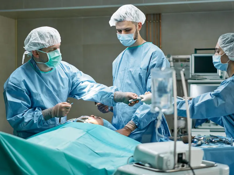
Treatment options
Treatment is planned according to the tumour site, stage, HPV status, the person's general health and their preferences. In early-stage tumours a single method (surgery or radiotherapy) is often enough, while advanced disease usually needs a combination.
Surgery
- Transoral surgery: selected laryngeal and pharyngeal tumours can be removed through the mouth without an external incision, under a microscope or endoscope, with laser or robotic systems; robotic surgery is one option for selected oropharyngeal tumours.
- Open resection: the tumour is removed with a margin of healthy tissue; when needed, part of the tongue or jawbone, or part or all of the larynx (partial or total laryngectomy), is removed.
- Neck dissection: removal of the lymph nodes in the neck, done to treat involved nodes or electively when the risk of spread is high.
- Reconstruction: when a large amount of tissue is removed, tissue transfer (a flap) is usually planned in the same operation to preserve swallowing, speech and appearance.
Radiotherapy and chemoradiotherapy
- Radiotherapy is used alone for some early tumours, as adjuvant treatment after surgery when there are high-risk features, or to preserve an organ; it is usually given as daily sessions over several weeks.
- Chemoradiotherapy: chemotherapy is given at the same time as radiotherapy in advanced disease, and is also used in larynx-preservation strategies.
- Techniques such as intensity-modulated radiotherapy (IMRT) aim to spare healthy structures such as the salivary glands and swallowing muscles.
Systemic therapy
- In recurrent or metastatic disease, immunotherapy (immune checkpoint inhibitors), chemotherapy and targeted drugs may be used alone or in combination; eligibility depends on tumour characteristics and laboratory tests.
Before and after surgery
Before surgery, an anaesthetic assessment, blood tests and, when needed, heart and lung assessment are carried out. Blood thinners and other regular medicines are adjusted only on the advice of the treating physician; do not stop them on your own. Stopping smoking helps wound healing and response to treatment.
- A dental assessment is important if radiotherapy is planned: the NCI advises a dental check at least a month before cancer treatment so the mouth can heal after any necessary dental work, because jawbone healing problems (osteoradionecrosis) can develop after radiotherapy.
- Nutritional status is assessed at diagnosis; dietitian support and a temporary feeding tube are planned when needed.
- A speech and swallowing therapist assesses the patient before treatment, and swallowing exercises may be recommended during radiotherapy.
- After major surgery a temporary tracheostomy (breathing opening in the neck) and a feeding tube may be needed; the hospital stay ranges from a few days to a few weeks depending on the operation.
- Pain is usually controlled with medicines; the team explains mouth, wound and drain care.
- Breathlessness, rapidly increasing neck swelling, bleeding, high fever, or redness or discharge from the wound should be reported immediately.
Recovery time and return to daily life vary with the extent of the operation and any additional treatment.
Follow-up
After treatment, regular check-ups aim to detect recurrence or a new second tumour early and to manage the long-term side effects of treatment.
- The American Cancer Society (ACS) guideline, endorsed by ASCO, recommends examinations every 1–3 months in the first year, every 2–6 months in the second year, every 4–8 months in years 3–5 and once a year after five years; the schedule is tailored to each person.
- Check-ups include examination of the mouth and neck and endoscopy, with imaging when needed; NICE recommends that, if PET-CT is done after chemoradiotherapy, it should be performed 3–6 months after treatment ends.
- Thyroid function is monitored in people whose neck was irradiated; the ACS guideline recommends measuring TSH every 6–12 months.
- Regular dental check-ups are needed, and dry mouth, swallowing, voice, nutrition, neck and shoulder movement, lymphoedema and mood are monitored.
- Stopping smoking and alcohol is part of follow-up; NICE stresses that continuing to smoke increases side effects, recurrence and the risk of a second cancer, and recommends offering help to quit.
If a new complaint appears between check-ups, seek review without waiting for the scheduled appointment.
Frequently asked questions
Sources
The general information on this page is based on the guideline and institutional sources below. Individual diagnosis and treatment decisions require an examination.
- NCI — Head and Neck Cancers fact sheet
- NCI — Oropharyngeal Cancer Treatment (PDQ, health professional)
- NICE NG36 — Cancer of the upper aerodigestive tract: assessment and management
- Cohen et al. — ACS Head and Neck Cancer Survivorship Care Guideline (2016)
- NCI — Oral complications of cancer therapies (PDQ)
- NHS — Mouth cancer symptoms