
General education only; not a substitute for examination or diagnosis.
What this area covers
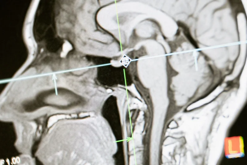
The skull base is the bony floor on which the brain rests, bordering the nose, sinuses, eye sockets, ears and neck. Nerves for vision, smell, hearing, facial movement and swallowing, and the large arteries supplying the brain, pass through its channels. The anterior skull base forms the roof of the nose and sinuses and the smell area; the central region contains the pituitary gland, the crossing of the optic nerves and the carotid artery; the lateral skull base includes the temporal bone, the internal auditory canal, the hearing and balance nerves and the facial nerve.
Conditions in this region are rare and varied; most are benign, but because they lie close to critical structures they need a team approach combining ENT–head and neck surgery, neurosurgery, endocrinology, ophthalmology, radiology and radiation oncology. The main conditions are:
- Pituitary adenoma: a usually benign tumour of the pituitary gland that can cause hormone excess or deficiency and narrowing of the visual field.
- Craniopharyngioma: a benign tumour arising near the pituitary stalk that is difficult to treat because of its location.
- Meningioma: a usually benign, slow-growing tumour arising from the membranes covering the brain.
- Chordoma and chondrosarcoma: rare, slow-growing tumours of skull base bone such as the clivus that tend to invade locally.
- Olfactory neuroblastoma (esthesioneuroblastoma): a rare malignant tumour arising from the smell area in the roof of the nose.
- Juvenile nasopharyngeal angiofibroma: a benign but highly vascular tumour, mostly in adolescent males, presenting with one-sided blockage and recurrent nosebleeds.
- Cerebrospinal fluid (CSF) leak and encephalocele: fluid or brain tissue protruding into the nose through a defect in the skull base; it may occur spontaneously or after trauma or surgery.
- Vestibular schwannoma (acoustic neuroma): a benign tumour of the hearing and balance nerve, among the most common lateral skull base tumours.
Symptoms
- Loss of smell, one-sided nasal blockage and recurrent nosebleeds.
- Narrowing of the visual field, especially at the outer edges, reduced vision or double vision.
- Hormonal symptoms: irregular periods, breast milk discharge, infertility, enlargement of the hands, feet or facial features, weight gain or persistent fatigue.
- One-sided hearing loss, ringing in the ear (tinnitus) and imbalance.
- Facial numbness or weakness, difficulty swallowing and voice change.
- Clear, watery discharge from one nostril that increases when bending forward, which suggests a CSF leak.
- Persistent headache or headache that changes in character.
Most of these symptoms have far more common, simple causes, but when they are one-sided and progressive they need assessment.
When to see a doctor
Situations that need emergency care
In the following situations go to an emergency department without delay or call 112 (the emergency number in Türkiye):
- Sudden, very severe headache, especially with loss of vision or double vision; emergencies such as sudden bleeding into the pituitary can present this way.
- Fever, neck stiffness and confusion; meningitis must be considered, especially in someone with watery nasal discharge.
- Sudden loss of vision, facial drooping, difficulty speaking or weakness of an arm or leg.
Situations that need a planned appointment
- Clear watery discharge from one nostril; because of the risk of meningitis it should be assessed promptly.
- One-sided, progressive hearing loss, tinnitus or imbalance.
- Narrowing of the visual field or unexplained hormonal symptoms.
- Loss of smell together with one-sided blockage or bleeding.
- A skull base mass found incidentally on imaging done for another reason.
This information is for general education. A definite diagnosis and treatment decision is made after examination, imaging and team review.
How it is diagnosed
- History and examination: detailed testing of the cranial nerves and endoscopic examination of the nasal cavity.
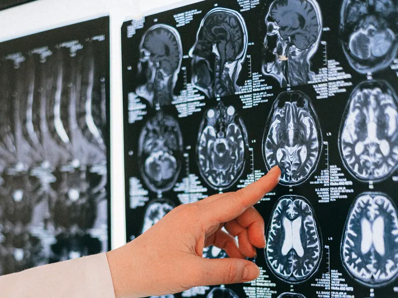
- Contrast-enhanced MRI: the main imaging test, showing how a lesion relates to soft tissue, nerves and the brain; it is the most valuable test for vestibular schwannoma and pituitary tumours.
- Computed tomography (CT): shows bone anatomy and erosion and is used for surgical planning and navigation.
- Angiography (CT, MR or catheter angiography): shows the relationship to major vessels; in highly vascular tumours such as angiofibroma it may also be used to block feeding vessels before surgery (embolisation).
- Endocrine tests: hormone levels in blood, and urine if needed, are measured for pituitary lesions.
- Eye examination and visual field testing: to assess pressure on the optic nerves.
- Hearing and balance tests: for lateral skull base lesions, tests such as audiometry and auditory brainstem response (ABR).
- When a CSF leak is suspected, the fluid is tested for beta-2 transferrin and thin-slice CT is used to locate the leak.
- Biopsy: performed when the tumour type would change treatment; it is generally avoided in highly vascular tumours because of bleeding risk.
Treatment options
Treatment is decided jointly by the team according to tumour type, size, growth rate, symptoms and the person's general health.
Observation and medical treatment
- Small, non-growing, symptom-free meningiomas, vestibular schwannomas or non-functioning pituitary adenomas can be monitored with regular MRI.
- For prolactin-secreting pituitary tumours (prolactinomas), dopamine agonist medicines are usually first-line; medicines are also used for other hormone-secreting tumours and for hormone deficiencies.
Surgical treatment
- Endoscopic endonasal surgery: an approach through the nose with a camera, without facial incisions; it can be used for pituitary adenomas, selected anterior and central skull base tumours and repair of CSF leaks. In most centres ENT and neurosurgical teams operate together.
- Open and combined approaches: if a tumour is too extensive to reach safely endoscopically, an approach through the skull (craniotomy), through the face, or a combination may be chosen.
- Lateral skull base surgery: for tumours such as vestibular schwannoma, the approach depends on tumour size and the level of hearing.
- Skull base reconstruction: the defect is closed in layers; the nasoseptal flap, a flap of septal lining with its own blood supply, is one widely used method.
Radiotherapy and other treatments
- Stereotactic radiosurgery and other forms of radiotherapy may be used for residual or recurrent tumour, or when surgical risk is high.
- For malignant tumours such as olfactory neuroblastoma, surgery, radiotherapy and, where needed, chemotherapy are planned by a multidisciplinary board.
Before and after surgery
- Before surgery, imaging, hormone and eye assessments and an anaesthetic review are completed. Blood thinners are changed only on the advice of the doctor who manages them.
- Possible risks include CSF leak, meningitis, bleeding, reduced smell, nasal crusting, hormone changes (for example diabetes insipidus, with excessive thirst and urination), and effects on vision or other nerves; risks depend on the tumour location and extent of surgery.
- After endoscopic surgery you will usually be asked not to blow your nose, to sneeze with your mouth open, and to avoid straining, heavy lifting and bending forward; your team will tell you how long these restrictions last.
- Saline spray or rinses usually start when your team advises; crusting is common and is cleared endoscopically at follow-up. When a nasoseptal flap has been used, crusting and smell change can last weeks to months.
- When to restart CPAP and when pressure changes such as air travel are allowed depend on the operation and should be discussed with your team.
- Seek care immediately for one-sided watery discharge that increases on bending forward or salty-tasting fluid running into the throat, severe headache, fever, neck stiffness, change in vision, excessive thirst and urination, or a nosebleed.
Follow-up
- The frequency of follow-up MRI depends on tumour type and treatment. Because tumours such as chordoma, meningioma and craniopharyngioma can recur years later, follow-up is usually long term.
- After pituitary surgery, hormone levels are monitored with endocrinology; hormone replacement is adjusted if needed and visual fields are rechecked.
- Healing inside the nose, crusting and the flap are assessed with nasal endoscopy.
- After lateral skull base treatment, hearing, balance and facial nerve function are monitored.
Frequently asked questions
Sources
The general information on this page is based on the guideline and institutional sources below. Individual diagnosis and treatment decisions require an examination.
- NCI — Pituitary tumor treatment
- AANS — Pituitary Gland and Pituitary Tumors
- NIDCD — Vestibular schwannoma (acoustic neuroma) and neurofibromatosis
- Hadad et al. — Vascular pedicle nasoseptal flap for skull base reconstruction (Laryngoscope, 2006)
- American Rhinologic Society — Postoperative precautions after endoscopic skull base surgery, expert practice statement (2024)